


-
SILICON VALLEY: Indian-origin executive named CEO of Microsoft Gaming - March 5, 2026
-
WASHINGTON: Indian-American lawyer at center of Trump’s biggest legal setback - March 4, 2026
-
TEXAS: ’15 of my cousins came here on H-1B’ - March 3, 2026
-
NEW YORK: Indian-origin doctor shares mother’s immigrant success journey in US - March 2, 2026
-
ARIZONA: Indian-origin scientist wins Arizona State University’s top Science Prize - March 1, 2026
-
WASHINGTON: Balaji Krishnamoorthy becoming Uber CFO amid ongoing visa row - February 28, 2026
-
LUCKNOW: Prime Minister Narendra Modi on HCL-Foxconn chip facility in UP - February 27, 2026
-
WASHINGTON: 55% Indian Americans Disapprove Of Trump’s India Policies: Survey - February 26, 2026
-
WASHINGTON: Trump Praises Indian American Harmeet Dhillon Amid Harvard Case - February 26, 2026
-
MUMBAI: Ranbir Kapoor to set up new RK Studios - February 25, 2026


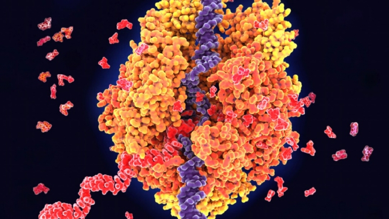
PARIS : 50 years ago, genes eluded electron microscopes
PARIS : Scientists still can’t directly see genes with electron microscopes, but combining the tool with the molecular scissors CRISPR/Cas9 has let researchers visualize genes being transcribed (illustrated) from DNA (blue) into RNA (red).
Molecular biologists can now visualize the larger structures of the cell, such as the nucleus and chromosomes, under the powerful electron microscope. But they have not been able to obtain images of genes (DNA) on the chromosomes. Nor have they been able to see RNA … or the intricate details of cell membranes, enzymes and viruses.
Update
Electron microscopes have become much more powerful over the last 50 years. For instance, in 1981, biophysicist Jacques Dubochet discovered that tiny biological structures supercooled with ethane could be observed in their natural state under an electron microscope. That finding paved the way for cryo-electron microscopy, which scientists use to visualize proteins, viruses and bacteria at the molecular level. Capturing detailed images of genes remains elusive, but scientists are inching closer. In 2021, researchers reported using an electron microscope and the molecular scissors CRISPR/Cas9 to visualize proteins transcribing DNA instructions for two genes into RNA.
About the Author
Some Related Posts
-
PARIS : NASA Hubble Spots Protective Shield Defending 2 Small Galaxies
PARIS : We may finally know how the Large and Small Magellanic Clouds are m -

PARIS : Sun’s Upcoming Peak of Sunspot and Solar Flare Activity Could Set Records
PARIS : The current solar cycle is intensifying with the expected peak of t -
PARIS: Gravitational wave ‘radar’ could help map the invisible universe
PARIS: It sounds like the setup for a joke: If radio waves give you radar a -

PARIS: Great white sharks may have helped drive megalodons to extinction
PARIS: For millions of years, megalodon sharks were top ocean predators — -

PARIS: Scientists Spot New Type of Stellar Explosion That’s Small But Fierce
PARIS: Astronomers have spotted a new type of thermonuclear explosion on th